
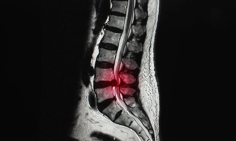
Back and neck pain often behave differently depending on posture. Many people feel worse after standing in line, walking through a store, or sitting at a desk for hours, yet feel noticeable relief the moment they lie down. That pattern is a clue about how the spine works, and it also explains a frustrating experience shared by many patients.
Someone with real, persistent symptoms has an MRI, and the report comes back showing little or nothing that explains the pain. In some of these cases, the problem is not missing. It simply was not visible in the position the scan was taken in.
How a Standard MRI Captures the Spine
Most MRI scans are performed with the patient lying flat on their back inside the scanner. This position works well for many purposes. It is comfortable for short periods, keeps the body still, and produces clear images of discs, nerves, bones, and soft tissue.
Lying down also takes the load off the spine. The vertebrae, discs, and ligaments are no longer supporting the weight of the head and torso, and the natural curves of the back relax. For conditions that are present in every position, this makes no difference. For conditions that depend on load, it can make a significant one.
What Changes When the Spine Carries Weight
Standing and sitting place the full weight of the upper body on the spine. Discs compress slightly, the spaces where nerves exit the spinal canal can narrow, and vertebrae that are not firmly held in place may shift. Bending forward or leaning back changes these relationships even more.
A weight-bearing MRI, sometimes called an upright or positional MRI, images the spine while the patient sits or stands. Some scanners also allow imaging in flexion and extension, meaning bent forward or leaning back, which shows how the spine moves through positions that reproduce symptoms in daily life.
Spine Conditions That Can Depend on Position
Spondylolisthesis is one of the clearest examples. In this condition, one vertebra slips forward over the one below it. When the slip is unstable, it may increase noticeably under load and partly reduce when the person lies flat, so a supine scan can understate how far the bone actually moves.
Foraminal narrowing is another. The foramina are the small openings where spinal nerves leave the canal, and they tend to get tighter when the spine extends or carries weight. A nerve that has enough room on a lying-down scan may be compressed when the person stands, which can explain leg or arm symptoms that otherwise seem unexplained.
Disc bulges and herniations can also look different under load. A bulge that appears mild in the supine position may press further toward the spinal canal or a nerve root when the disc is compressed by body weight.
In the neck, instability of the upper cervical spine and the relationship between the skull and the top vertebrae are known to change with position. Patients evaluated for craniocervical instability or Chiari malformation are sometimes imaged upright because the findings that matter most can depend on how the head and neck are positioned.
Signs a Positional Scan May Be Worth Discussing
A weight-bearing study is not needed for every back problem, and a standard MRI remains the right first step for many patients. Still, certain patterns suggest the question is worth raising with a physician.
These include pain that clearly worsens with standing, sitting, or walking and eases when lying down, symptoms that do not match a previous normal or mild MRI, sciatica that appears only in certain postures, and suspected instability after an injury or prior surgery. People evaluating options for MRI of the back and spine often bring these exact patterns to their appointments.
Comfort and Practical Benefits
Upright scanners usually have an open design, with no enclosed tube surrounding the body. Many patients who feel anxious or claustrophobic in conventional machines find this setup much easier to tolerate, and some can watch a screen during the exam.
Being able to sit or stand also helps patients who cannot lie flat because of pain, breathing difficulty, or body size. A scan that a patient can complete without moving tends to produce clearer, more useful images.
Understanding the Trade-Offs
Positional MRI is a tool for specific questions rather than a replacement for every standard scan. Many upright systems use lower magnetic field strengths than high-field closed scanners, which can mean longer scan times and differences in image detail for some types of studies.
For that reason, physicians often decide based on what they are trying to answer. When the main question is whether something changes with load or movement, a weight-bearing scan may offer information that a supine study cannot. When the question is about fine detail in a part of the body where position does not matter, a conventional scan may be the better fit.
Talking With Your Doctor About Imaging Options
Patients get the most from any MRI when the ordering physician understands their symptoms in detail. Describing exactly which positions make pain better or worse, how long symptoms last, and what activities trigger them helps the physician choose the right study.
If you already had a standard MRI that did not explain your symptoms, bring the images and report to your follow-up visit. A comparison between supine and weight-bearing images can sometimes clarify a diagnosis that has stayed uncertain for months.
Conclusion
The spine is designed to carry weight, and some of its problems only reveal themselves when it does. Slipping vertebrae, narrowing nerve openings, and load-dependent disc changes can be understated on a scan taken lying down.
For patients whose symptoms clearly follow posture, imaging the spine upright can add useful information, and conditions such as craniocervical instability evaluated with positional MRI show how much position can change what a scan reveals.